Micro-CT and layered tektites
Decoding Muong Nong: What Micro-CT Scans Reveal Inside Layered Tektites
A Muong Nong specimen can look banded, blocky, swirled, or bubble-rich on the outside. Micro-CT asks a narrower question: does that visible pattern continue into the interior as a real 3D structure? For the Muong Nong layered structure, micro-CT scans can reveal internal vesicles, elongated voids, inclusions, and uneven layer relationships without cutting the tektite open. The limit matters just as much: a scan can document internal geometry, but it does not by itself establish the full formation sequence, melt chemistry, authenticity, temperature history, or every stage of plastic deformation.
broader context
Start with the main moldavite page
This narrower page makes more sense after the broader moldavite archive page.
What Micro-CT Adds
Collector language usually starts at the surface. Muong Nong-type pieces are often described as layered, banded, swirled, blocky, or less uniform than splash-form tektites. Those words are useful because they describe what the eye notices first. They are not enough to settle the structural question.
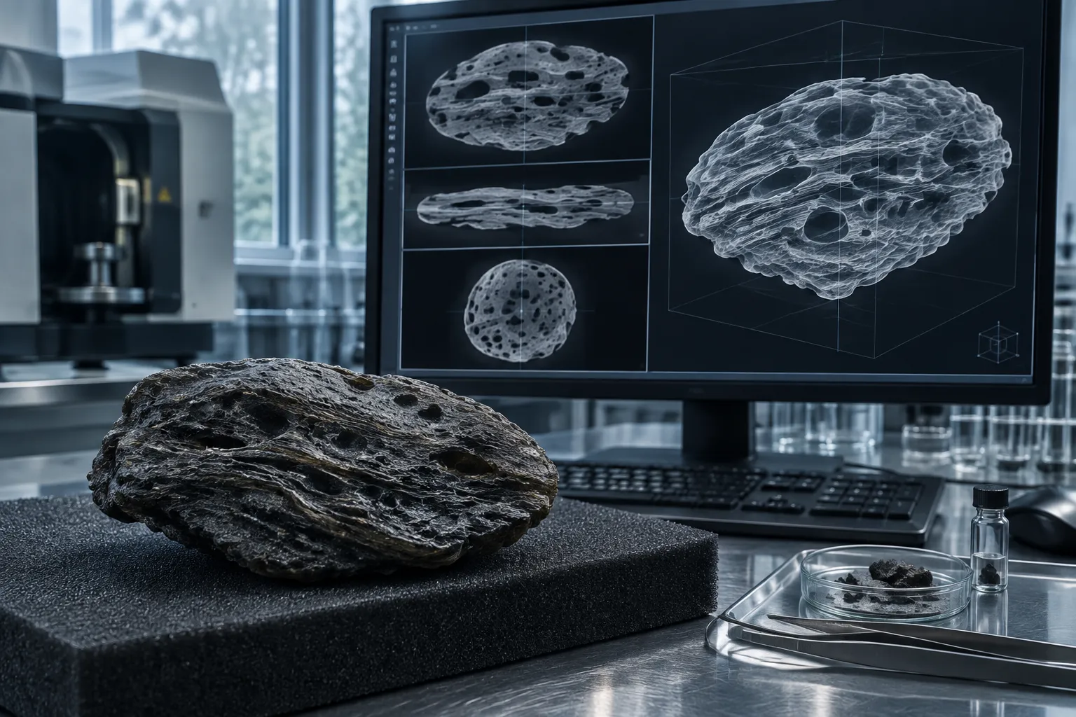
Micro-CT shifts the problem from a surface reading to a volume reading. Instead of relying only on broken edges, polished windows, or exterior bands, an X-ray micro-CT scan reconstructs interior slices into a 3D model. In that model, voids, denser inclusions, angular fragments, and layer-related heterogeneity can be mapped in relation to one another.
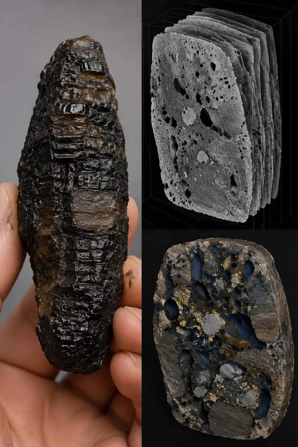
That is the value for Muong Nong-type tektites. Their layered character is not just a decorative surface feature in the scientific literature. They are described as irregular, blocky, and internally heterogeneous, and tomography adds a second view: some examined pieces contain internal voids, elongated vesicles, inclusions, and fragment-like relationships that are not visible from the outside alone.
The scan does not make the specimen more dramatic. It makes the claim more specific.
Careful reading
This scan shows voids and inclusions arranged within a heterogeneous layered tektite volume.
Careless reading
This scan settles exactly how all Muong Nong tektites formed.
The first fits the evidence. The second goes beyond it.
Vesicles, Inclusions, and Layer Heterogeneity
The most useful features in Muong Nong micro-CT scans are usually vesicles, inclusions, and changes in internal texture from one part of the specimen to another.
A vesicle is a gas bubble preserved as a void in the glass. In collector language, these are the bubbles. In scan language, they become measurable void spaces, subject to the resolution and processing limits of the image. Some CT observations of Muong Nong-type material report elongated vesicles rather than only round bubbles. Elongation can matter because it records directionality in the preserved glass, but it is not a full explanation by itself.
An elongated vesicle may suggest that the material was stretched, compressed, or otherwise deformed while still able to change shape. That is why plastic deformation appears in discussions of Muong Nong structure. But elongated vesicles in tektites do not, on their own, establish a single exact deformation path. They need to be read alongside layering, fragment geometry, mineral inclusions, and broader formation models.
Inclusions are another important scan target. X-ray tomography has been used in research on Muong Nong-type material to examine the spatial setting of mineral inclusions, including work connected with coesite preservation. That kind of scan can show where an inclusion sits inside the glass and how it relates to nearby voids or internal structures. It does not automatically identify every chemical detail. For that, compositional or mineralogical analysis is needed.
Layer heterogeneity is the broader pattern. A scanned specimen may show that voids are not evenly distributed, that one zone differs from another, or that apparent layers have different internal textures. Older geochemical and morphological work describes Muong Nong-type tektites as layered and blocky, with bubble abundance differing across contrasting layers. The exact comparisons belong to the source material, not to a blanket claim about every specimen. Still, the direction is clear: the layers are treated as structural differences, not only surface decoration.
For collectors, the practical translation is simple. Surface bands, grooves, and swirls may be a clue. Internal layering is a stronger claim. Micro-CT helps test the gap between those two.
Porosity Mapping Is Processed Evidence
Micro-CT porosity mapping is often shown as a bright, colored 3D model. It can look like a direct image of the inside of the stone. It is not that simple.
A porosity map is processed output. The scan records X-ray contrast through the specimen. Software reconstructs slices, then analysts choose thresholds and segmentation rules that separate glass from voids or other features. The final map depends on voxel size, scan quality, reconstruction choices, specimen boundaries, and the selected volume of interest.
That does not make porosity mapping unreliable. It makes it conditional.
For Muong Nong porosity mapping, the strongest use is descriptive and spatial: where are the vesicles, how large are the visible voids within the scan limits, are they isolated or connected, and do they vary from one layer-like zone to another? Those are legitimate structural-analysis questions. They can help distinguish a more homogeneous-looking glass volume from a more internally complex specimen.
The weaker use is importing numbers from elsewhere. Porosity values from moldavite, bricks, carbonates, sandstones, tissue scaffolds, or vendor sample scans should not be assigned to Muong Nong-type tektites. Even moldavite data require caution here. Moldavites are tektites, but they are not Muong Nong-type Australasian layered tektites. They belong to a different strewn field.
That distinction matters because the page slug includes “moldavite,” and collecting-market language sometimes blurs tektite categories. A Muong Nong-type tektite should not be called moldavite. Micro-CT methods used on tektite glass can inform vocabulary and workflow, but moldavite porosity measurements are not Muong Nong measurements.
Open and closed porosity also need careful handling. “Open” and “closed” refer to connectivity, not simply big versus small bubbles. A small void might connect to a larger network; a larger bubble might be isolated. A 2D slice can suggest patterns, but stronger connectivity claims need the 3D volume and careful segmentation.
This is where micro-CT is useful and modest at the same time. It can make hidden geometry visible. It cannot remove the need for interpretation.
What the Scans Suggest About Formation
The most current formation language around Muong Nong layering often points toward incomplete melt mixing. In plain terms, layers may reflect melts or melt portions with slightly different properties that did not fully homogenize before the glass was preserved. That fits the way Muong Nong-type tektites are discussed as layered, heterogeneous impact glass rather than uniformly mixed splash forms.
Micro-CT can support that discussion by showing internal heterogeneity. It can show vesicle orientation, fragment relationships, inclusions, and differences between zones. It can make a layered tektite structure easier to inspect without slicing the specimen apart.
But micro-CT does not directly establish incomplete melt mixing. It does not identify melt chemistry by itself. It does not give a full temperature history. It does not reconstruct the flight path, the exact timing of deformation, or the order in which every layer formed. If a scan shows elongated vesicles, that observation can be consistent with deformation, but it should not be inflated into a complete plastic-deformation reconstruction.
That boundary matters because Muong Nong specimens invite storytelling. Their blocky shapes, layered interiors, and uneven textures can make them feel more explained than the evidence allows. A broken edge might suggest layering. A surface band might suggest flow. A scan might reveal elongated vesicles. Each point adds information, but none of them alone closes the formation question.
A more careful reading is layered, not absolute
- The exterior may show bands, swirls, grooves, or blocky form.
- The scan may show internal vesicles, inclusions, and heterogeneous zones.
- Formation models may interpret the layering through incomplete melt mixing and deformation.
- Additional mineralogical and compositional work is needed before stronger claims are made.
That keeps specimen traits before the story.
Surface Banding Versus Internal Layering
One common confusion is treating every visible band as proof of internal layering. Exterior texture can exaggerate, obscure, or only partly reflect what is happening inside a tektite. Weathering, fracture surfaces, natural edges, and lighting all change what a collector sees.
Micro-CT helps because it does not rely on the exposed surface alone. If the scan shows layer-related changes in void distribution, internal streaks, inclusions, or textural variation through the volume, the structural case becomes stronger. If the scan shows mostly superficial contrast with little internal continuation, then the visible surface pattern should be read more cautiously.
The opposite mistake is also possible: dismissing collector observations because they start with surface language. Words like “bands,” “swirls,” and “bubbles” are not measurements, but they are often the starting point for a real question. The edit step is to translate them. “Bubbles” become vesicles. “Bands” become possible layer relationships. “Swirls” become possible internal heterogeneity or flow-like texture, depending on what the scan and supporting analyses actually show.
A scan can also complicate the visible impression. A specimen that looks quiet outside may contain internal voids or inclusions. A dramatic surface may not carry the same pattern through the interior. That is why micro-CT is useful for structural analysis; it tests the inside, not just the look.
Limits That Matter
Micro-CT scan limitations are not fine print. They shape what can be said.
Resolution
Features smaller than the effective voxel size may be missed or blurred. A scan that captures larger vesicles may not resolve fine pores, hairline cracks, or tiny inclusions. Without scan parameters, it is not responsible to claim a complete pore-size distribution.
Segmentation
Turning grayscale scan data into a void map requires threshold choices. Different thresholds can change apparent porosity, especially in complex glass with inclusions, artifacts, or weak contrast. Ring artifacts and reconstruction issues can also interfere with interpretation if they are not controlled.
Specimen selection
A single Muong Nong-type tektite cannot define the whole class. Even within one piece, the selected volume of interest can change the apparent pattern. A scan through one zone may show more vesicles than another. A broken fragment may not represent the original whole.
Collector questions
Micro-CT is not an authenticity verdict. It may reveal internal features consistent with natural tektite structure, but authenticity also depends on provenance, external morphology, composition, and comparison with known material.
Nor does micro-CT support claims about value, symbolic effect, or personal transformation. Those are separate interpretive or market conversations, and they should not be folded into the scan result.
The scan answers a structural question. It does not answer every collector question.
Practical Reading
The most defensible reading of Muong Nong micro-CT scans is this: they reveal the hidden 3D arrangement of vesicles, inclusions, fragments, and heterogeneous layers inside examined layered Australasian tektites. They can strengthen the distinction between surface banding and internal layering. They can support cautious discussion of incomplete melt mixing and possible plastic deformation when paired with other evidence.
They cannot establish a universal formation sequence, assign porosity values without scan-specific methods, transfer moldavite measurements to Muong Nong material, or authenticate a specimen on their own.
For a collector or moldavite reader trying to place Muong Nong material correctly, that is the useful boundary. Muong Nong-type tektites are not moldavites, but they belong to the wider tektite conversation where impact glass, internal structure, and provenance all matter. Micro-CT adds a strong interior view. The interpretation still has to stay inside what the scan actually shows.